Clinical significance of FLT3-ITD in Acute Myeloid Leukemia
FLT3 is a class III receptor tyrosine kinase expressed mainly on hematopoietic progenitor cells, where its juxtamembrane (JM) domain holds the kinase inactive until ligand binding (Figure 1) [1]. FLT3 internal tandem duplication (FLT3-ITD) is an in-frame, head-to-tail duplication of a segment within the JM domain or the adjacent first tyrosine kinase domain (TKD1) [2]. It disrupts JM autoinhibition, so the receptor signals constitutively without ligand [3]. FLT3-ITD occurs in about 25% of AML. It is associated with high leukemic burden, high relapse rate and short overall survival, and because it can appear or disappear through clonal evolution, testing is recommended at both diagnosis and relapse [4].
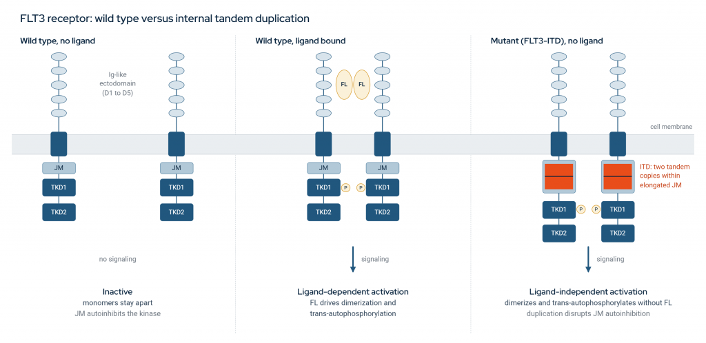
Figure 1: FLT3 receptor: wild type versus internal tandem duplication. In the absence of ligand, wild-type FLT3 remains monomeric and the juxtamembrane (JM) domain keeps the kinase inactive (left). FLT3 ligand (FL) drives dimerization and trans-autophosphorylation (P), activating downstream signaling (middle). The internal tandem duplication in the JM domain disrupts autoinhibition, so the mutant receptor dimerizes and signals without ligand (right). Ig, immunoglobulin-like domains (D1 to D5); TKD1/TKD2, split kinase domain.
Beyond its presence or absence, the proportion of FLT3 alleles carrying the ITD also matters. This proportion is expressed as the allelic ratio (AR): the ITD signal divided by the wild-type signal. The 2017 ELN recommendations combined AR with NPM1 status to assign risk. Patients with mutated NPM1 and a low AR (below 0.5) were classed as favorable, whereas a high AR (0.5 or above) placed them in the intermediate group, or in the adverse group if NPM1 was wild type [5]. The AR value could therefore change a patient’s risk category and, with it, whether transplant was considered. ELN 2022 removed AR from risk classification because current assays are not reproducible [6]. Accurate quantification still matters, however, for measurable residual disease (MRD) monitoring [7] and for selecting patients for FLT3 inhibitors such as midostaurin, gilteritinib and quizartinib [4].
PCR followed by fragment analysis is the clinical reference standard, with AR calculated from the ITD and wild-type peak intensities [8]. Fragment analysis does not reveal the duplicated sequence or the insertion site, whereas NGS can report both while testing other AML genes in the same assay [9].
Why FLT3-ITD is hard to call from short reads
An ITD duplicates sequence already present in the reference, so the aligner has no novel anchor (Figure 2). Duplications also vary in length (median 48 bp, range 15 to 180) and in insertion site [2, 10], and this variation carries clinical information, as insertion site may be an independent adverse prognostic factor [2].
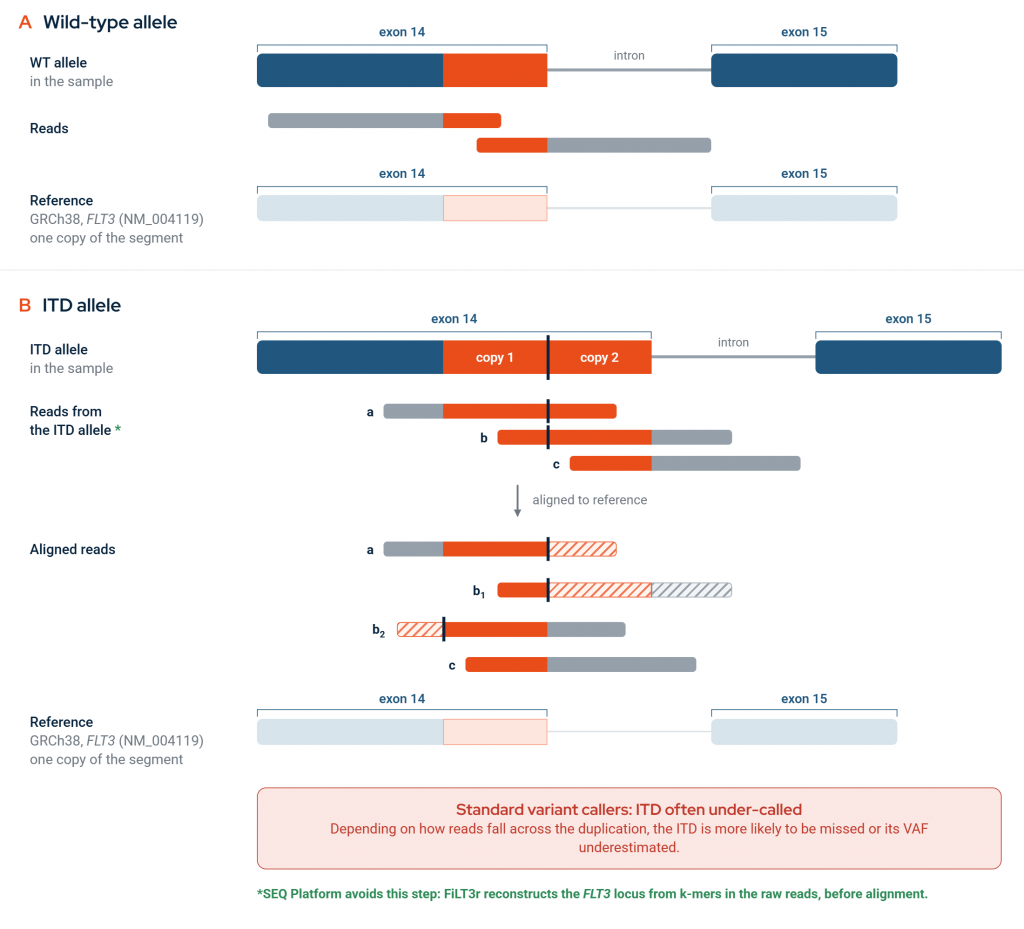
Figure 2: Why standard short-read alignment under-detects FLT3-ITD. (A) Reads from the wild-type (WT) allele align to the reference without discrepancy. (B) In an internal tandem duplication, the segment is present as two head-to-tail copies (copy 1 and copy 2). The black vertical line marks the duplication junction. Reads that cross the junction (a, b) contain a junction that is absent from the reference, which has room for only one copy. The aligner places the part of the read that matches and soft-clips the rest (hatched). The same read can be aligned in more than one way: read b is shown soft-clipped either after the junction (b1) or before it (b2). Reads that lie within a single copy (c) align cleanly and carry no evidence of the ITD. Depending on how reads fall across the duplication, standard variant callers may miss the ITD or underestimate its variant allele fraction (VAF). SEQ Platform does not rely on this alignment: FiLT3r reconstructs the FLT3 locus from k-mers in the raw reads, before alignment.
Standard short-read workflows handle this structure poorly (Figure 2). In a systematic analysis of 46 clinical cases, short-read alignment misaligned reads for every insertion and deletion examined, and the fraction of misaligned reads increased with variant size. As a result, variant allele fraction was undercalled by 1% to 84%, and in at least one case a FLT3-ITD detected by capillary electrophoresis was not reported [11]. The problem is greatest for large ITDs, and standard tools consistently underestimate allele burden unless soft-clipped and junctional reads are recovered [12]. Reliable detection therefore requires methods designed for the ITD structure: reassembling misaligned reads [11], recovering soft-clipped and junctional reads [12], or avoiding alignment entirely with k-mer analysis [13].
FLT3-ITD detection in SEQ Platform
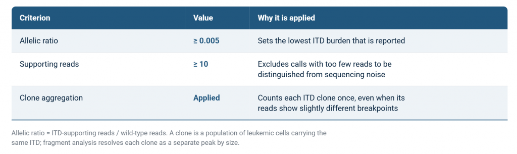
SEQ Platform detects FLT3-ITDs with FiLT3r [13]. Reads carrying an ITD frequently fail to align correctly, so FiLT3r does not rely on the aligned BAM file. Instead, it reconstructs the FLT3 locus from k-mers in the raw reads. Detection runs automatically and requires no separate assay or setting. It is limited to the ITD hotspot region, which covers FLT3 exons 14 and 15, where ITDs occur. A detected event is reported only when it meets all three criteria in Table 1. The minimum of 10 supporting reads removes low-depth read clusters that cannot be separated from background noise. Clone aggregation addresses a common pattern in which the same clone is observed with slightly different breakpoints: these observations are merged into a single clone, so the reported clone count reflects the clonal composition of the sample.
Table 1: Reporting criteria for FLT3-ITD events.
Reported ITDs are added to the small variant list as chromosome 13 insertions, with FiLT3r shown as the calling tool, and pass through the same annotation steps as other variants. They therefore appear in Clinical Evidence and Clinical Trials tabs wherever matching content exists, so ITD status is reviewed together with the rest of the sample’s variants.
Performance against fragment analysis
SEQ Platform was evaluated on the published FiLT3r evaluation cohort of 185 AML samples, with PCR fragment analysis (GeneScan) as the reference [13]. The cohort comprises 114 FLT3-ITD-positive and 71 FLT3-ITD-negative samples. The positive samples carry 147 truth clones: 83 shorter than 50 bp, 55 between 50 and 150 bp, and 9 longer than 150 bp.
Table 2: FLT3-ITD detection performance against PCR fragment analysis.
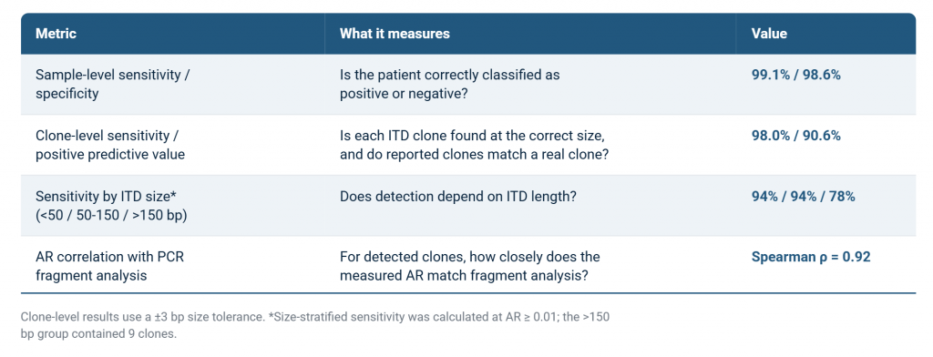
Table 2 addresses 3 questions. The first is whether the patient is correctly classified: of the 114 FLT3-ITD-positive samples, 113 were called positive (sample-level sensitivity 99.1%), and of the 71 negative samples, 70 were called negative (sample-level specificity 98.6%), so 183 of 185 samples were classified correctly. The second is whether each individual ITD is found. Some patients carry more than one ITD clone, and fragment analysis identified 147 clones in the 114 positive samples. SEQ Platform detected 98.0% of these at the correct size (within ±3 bp), and 90.6% of the clones it reported matched a clone seen by fragment analysis (positive predictive value). Sensitivity was 94% for ITDs shorter than 50 bp, 94% for those between 50 and 150 bp, and 78% for those longer than 150 bp, although this last group contained only 9 clones. The third is how accurately ITD burden is measured. For clones detected by both methods, the allelic ratio from SEQ Platform closely tracked the fragment analysis value (Spearman ρ = 0.92, where 1 indicates identical ranking). This correlation describes how precisely burden is measured, not how often ITDs are found.
Conclusion
FLT3-ITD status and burden inform prognosis and the choice of FLT3 inhibitors in AML, yet the structure of the duplication makes it difficult to detect with standard short-read pipelines. SEQ Platform addresses this within routine somatic analysis, reporting FLT3-ITDs alongside the rest of the sample’s variants and the matching clinical evidence and trials. On the benchmark cohort, it classified nearly all samples correctly, resolved individual clones in samples carrying more than one ITD, and produced allelic ratios that closely tracked fragment analysis. SEQ Platform provides FLT3-ITD status, clonal composition and allelic ratio from the same NGS run used for the rest of the AML workup, giving laboratories the information they need to support interpretation and treatment decisions.
References
- Naoe T, Kiyoi H. Oncogenic protein tyrosine kinases. Cell Mol Life Sci. 2004;61(23):2932-2938. doi:10.1007/s00018-004-4274-x
- Kayser S, Schlenk RF, Londono MC, Breitenbuecher F, Wittke K, Du J, et al. Insertion of FLT3 internal tandem duplication in the tyrosine kinase domain-1 is associated with resistance to chemotherapy and inferior outcome. Blood. 2009;114(12):2386-2392. doi:10.1182/blood-2009-03-209999
- Griffith J, Black J, Faerman C, Swenson L, Wynn M, Lu F, et al. The structural basis for autoinhibition of FLT3 by the juxtamembrane domain. Mol Cell. 2004;13(2):169-178. doi:10.1016/s1097-2765(03)00505-7
- Daver N, Schlenk RF, Russell NH, Levis MJ. Targeting FLT3 mutations in AML: review of current knowledge and evidence. Leukemia. 2019;33(2):299-312. doi:10.1038/s41375-018-0357-9
- Döhner H, Estey E, Grimwade D, Amadori S, Appelbaum FR, Büchner T, et al. Diagnosis and management of AML in adults: 2017 ELN recommendations from an international expert panel. Blood. 2017;129(4):424-447. doi:10.1182/blood-2016-08-733196
- Döhner H, Wei AH, Appelbaum FR, Craddock C, DiNardo CD, Dombret H, et al. Diagnosis and management of AML in adults: 2022 recommendations from an international expert panel on behalf of the ELN. Blood. 2022;140(12):1345-1377. doi:10.1182/blood.2022016867
- Blätte TJ, Schmalbrock LK, Skambraks S, Lux S, Cocciardi S, Dolnik A, et al. getITD for FLT3-ITD-based MRD monitoring in AML. Leukemia. 2019;33(10):2535-2539. doi:10.1038/s41375-019-0483-z
- He R, Devine DJ, Tu ZJ, Mai M, Chen D, Nguyen PL, et al. Hybridization capture-based next generation sequencing reliably detects FLT3 mutations and classifies FLT3-internal tandem duplication allelic ratio in acute myeloid leukemia: a comparative study to standard fragment analysis. Mod Pathol. 2020;33(3):334-343. doi:10.1038/s41379-019-0359-9
- Duncavage EJ, Tandon B. The utility of next-generation sequencing in diagnosis and monitoring of acute myeloid leukemia and myelodysplastic syndromes. Int J Lab Hematol. 2015;37 Suppl 1:115-121. doi:10.1111/ijlh.12361
- Blau O, Berenstein R, Sindram A, Blau IW. Molecular analysis of different FLT3-ITD mutations in acute myeloid leukemia. Leuk Lymphoma. 2013;54(1):145-152. doi:10.3109/10428194.2012.704999
- Craven KE, Fischer CG, Jiang LQ, Pallavajjala A, Lin MT, Eshleman JR. Optimizing insertion and deletion detection using next-generation sequencing in the clinical laboratory. J Mol Diagn. 2022;24(12):1217-1231. doi:10.1016/j.jmoldx.2022.08.006
- Tung JK, Suarez CJ, Chiang T, Zehnder JL, Stehr H. Accurate detection and quantification of FLT3 internal tandem duplications in clinical hybrid capture next-generation sequencing data. J Mol Diagn. 2021;23(10):1404-1413. doi:10.1016/j.jmoldx.2021.07.012
- Boudry A, Darmon S, Duployez N, Figeac M, Geffroy S, Bucci M, et al. Frugal alignment-free identification of FLT3-internal tandem duplications with FiLT3r. BMC Bioinformatics. 2022;23(1):448. doi:10.1186/s12859-022-04983-6