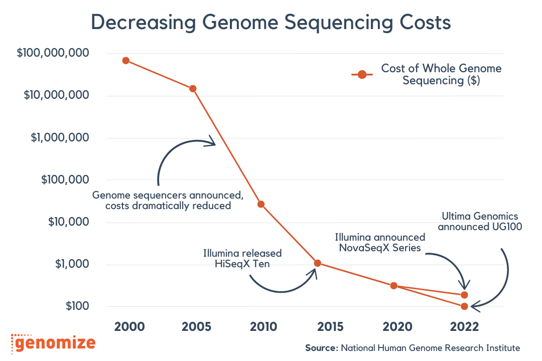
The sequencing of the human genome, together with steep drops in cost and turnaround time, has rapidly expanded what we know about the genomic alterations that drive cancer. This has given clinicians new tools to match care to the biology of an individual tumor, from genomically informed prognosis to targeted therapies. As in germline disease, next-generation sequencing (NGS) is increasingly favored over traditional single-target methods because it tests many genes at once and saves both time and cost (1).
Sequencing is only the first step. A variant call has to be interpreted before it means anything, and that is where germline and somatic analysis part ways. They run on the same sequencing technology, yet the question you ask of a variant, and the rules you use to classify it, are fundamentally different.
Germline vs. Somatic Variants
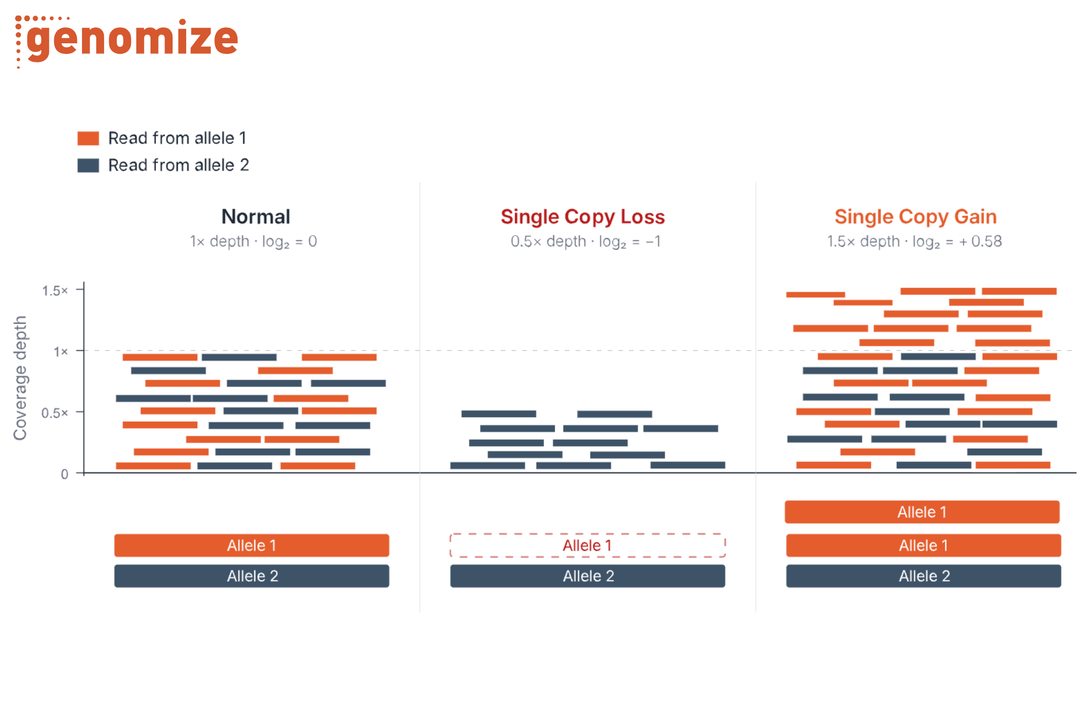
A germline variant is an alteration you are born with. It is present in every cell* of the body and is passed down from a parent or arises in the egg or sperm, which means it can be inherited. A somatic variant is an alteration a cell acquires during a person’s life. It is found only in the tumor and the cells that descend from it, it is not inherited, and it is often present in only a fraction of the sample (its variant allele fraction) (Figure 1). So the questions differ. For a germline variant, we want to know whether it causes or predisposes to inherited disease and with what likelihood (pathogenicity and penetrance). For a somatic variant, we want to know whether it is driving the cancer and whether it can be targeted with treatment (oncogenicity and actionability).
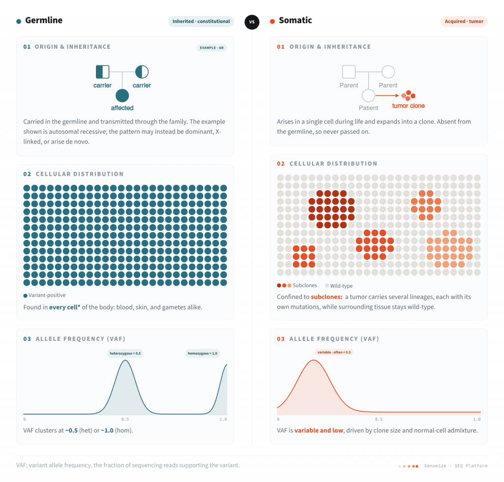
Figure 1. Germline versus somatic variants differ in origin and inheritance, cellular distribution, and observed allele frequency (VAF). Germline variants are present in every cell* and show VAF near 0.5 (heterozygous) or 1.0 (homozygous); somatic variants are confined to the tumor clone and typically show lower, more variable VAF shaped by clone size, tumor purity, and copy number. *With exceptions. Post-zygotic mosaic variants arise after fertilization and are carried by only a subset of cells.
Classifying A Somatic Variant: The Frameworks and The Knowledgebases Behind Them
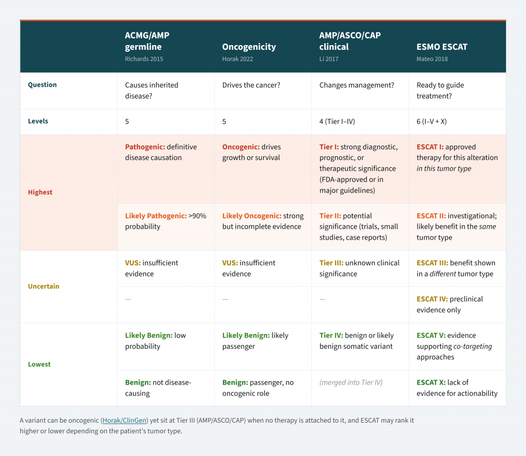
These different questions are answered by different formal frameworks. In 2015, Richards and colleagues proposed guidelines for interpreting sequence variants in genes linked to Mendelian disorders, setting the standard for deciding whether a variant causes inherited disease (2). Their framework sorts germline variants into five categories, from benign to pathogenic, weighing evidence such as population frequency, computational predictions, and functional data. This became the foundation for clinical variant interpretation. But cancer needs more than a single judgment about whether a variant causes disease, because a tumor raises two more questions, and each has its own framework.
The first is whether the variant is driving the cancer. A tumor genome carries many changes, but only some give the cell a growth or survival advantage. These drivers have to be separated from the far more numerous passengers that are simply carried along. To make that call, ClinGen, the CGC, and VICC published standards (3) that classify a variant’s oncogenicity, placing it on a five-level scale from benign to oncogenic using evidence codes adapted from the germline ACMG system.
The second question is whether the variant changes what the clinician does next. A variant can be a real driver and still have no treatment attached to it, so knowing it is oncogenic does not tell you how to act on it. The AMP, ASCO, and CAP guidelines (4) address this with a four-tier system that ranks variants by the strength of their clinical evidence, from Tier I, with strong diagnostic, prognostic, or therapeutic significance, down to Tier IV. ESMO’s ESCAT scale (5) works in the same spirit, ranking genomic alterations by how ready they are to guide treatment in a specific tumor type.
Table 1. Somatic and germline variant classification frameworks at a glance. ACMG/AMP and the oncogenicity framework classify a variant the same way regardless of tumor type. AMP/ASCO/CAP and ESCAT are both tumor-type-specific by design: the same variant can occupy different tiers in different cancers. ESCAT is an actionability ranking rather than a benign-to-oncogenic axis, so its lower tiers describe evidence strength, not a ‘benign’ call.
Every one of these frameworks runs on curated evidence, and in cancer that evidence lives in a few knowledgebases most labs come to rely on.
- OncoKB, maintained by Memorial Sloan Kettering, grades variant-drug relationships on its own levels of evidence and in 2021 became the first somatic database to earn partial FDA recognition (6, 7).
- CIViC takes an open, community-curated approach, with every interpretation traceable back to the literature and free for anyone to use (8).
- CKB, the Cancer Knowledgebase, works along the same lines and pairs each variant or molecular profile with the therapies, clinical evidence, and open trials attached to it, grades that evidence on the AMP/ASCO/CAP tiers, and traces each entry back to its guideline (NCCN, ESMO) or primary-literature source (9).
- COSMIC plays a different role. It is primarily a catalogue of which mutations have been seen across cancers, along with their frequencies and associated mutational signatures (10).
Oncogenicity vs. Actionability
Oncogenicity is largely a property of the variant. An alteration that disables a tumor suppressor or switches on an oncogene does so wherever it occurs, so its oncogenicity stays the same from one tumor to the next. Actionability depends on context: the tumor type, the drugs that exist, and the evidence behind them. A variant can therefore hold a fixed oncogenicity and still move between clinical tiers depending on where it shows up.
BRAF V600E shows this plainly. It is an oncogenic driver in both melanoma and colorectal cancer, and that classification does not change between them. What changes is its clinical tier which depends on the drug and the tumor type. In melanoma, V600E paired with a BRAF inhibitor is a Tier I, ready-to-use target, and the drug given alone produces strong responses (11). In colorectal cancer the same variant does not reach that tier for the same drug: a BRAF inhibitor alone works poorly, because blocking BRAF triggers feedback activation through EGFR that keeps the cell growing (12). The variant only becomes Tier I in colorectal cancer when paired with a different strategy, a BRAF inhibitor together with an anti-EGFR antibody (13). One variant, one oncogenicity, but the same drug earns a different tier in each tumor.
This is also why a clinical tier is a snapshot, not a fixed label. Oncogenicity reflects the biology of the variant and stays put; a tier reflects the evidence available at the time. That evidence grows, so the same variant can become more actionable over time without its biology changing at all.
What the Allele Fraction Tells You
The variant allele fraction is the share of sequencing reads that carry the change. In germline testing it barely moves: about half the reads for a heterozygous variant, nearly all for a homozygous one, so it confirms little you don’t already know. In a tumor it does real work. A high fraction points to a variant present in most of the cancer’s cells, and likely an early event; a low one points to a smaller subpopulation, and a later event (14).
Allele fraction is only as clean as the sample it comes from. It reads as a proportion of tumor cells only when the sample is nearly pure tumor at normal copy number, which it often is not. Low tumor purity drags every fraction down at once, so a clonal driver can look subclonal. Copy-number changes distort it locally too: an amplification or a loss of heterozygosity shifts the fraction of any variant sitting on the affected segment.
An allele fraction is read against the tumor’s purity and local copy number, never on its own.
The reads underneath the fraction have to be trustworthy as well. At low fractions a real variant becomes hard to separate from a PCR or a sequencing error, and this is where unique molecular identifiers (UMIs) earn their place. Each original DNA fragment is tagged before amplification, so duplicate reads collapse back to the single molecule they came from (15). UMIs therefore make confident calling possible at very low fractions which is typical for circulating tumor DNA and liquid biopsies (16).
How the sample was sequenced matters just as much. With a matched normal, the patient’s own germline variants can be subtracted directly, leaving a cleaner set of genuinely somatic calls. A tumor-only run has no such comparison, so it relies on population-frequency databases to infer which alterations are inherited, and some germline variants inevitably slip through disguised as somatic.
Sample-Level Signals: TMB and MSI
Alongside the question of whether a specific variant is oncogenic and actionable, some of the most consequential decisions in oncology rest on a property of the tumor as a whole. Tumor mutational burden (TMB) and microsatellite instability (MSI) are two such genome-scale biomarkers. Both are read out of the same sequencing data used for variant calling, and both tend to point toward immunotherapy rather than a targeted drug.
Tumor mutational burden (TMB) is, in essence, a count: the number of somatic mutations per megabase of tumor DNA (17). The biological logic is that a heavily mutated tumor produces more abnormal proteins, making it more visible to the immune system and more likely to respond to immune checkpoint inhibitors (18). This is the reasoning behind pembrolizumab’s approval for TMB-high (≥10 mutations/Mb) solid tumors. Thresholds are assay-dependent and the predictive value varies by tumor type.
Microsatellite instability (MSI) is a signature of a broken repair system. When a tumor’s DNA mismatch repair (MMR) machinery fails, short repeated motifs called microsatellites accumulate insertions and deletions, and the tumor reads as MSI-high. A tumor like this is effectively mismatch-repair deficient, and that status carries real weight. It was the basis of the first cancer treatment the FDA cleared on molecular grounds alone, regardless of where the tumor arose (19). High TMB and MSI-high often show up in the same tumor, but they are not the same measurement, and either one on its own can open the door to a checkpoint inhibitor.
SEQ Platform: From Variant Call to Clinical Decision
SEQ Platform is built around these distinctions. It separates somatic from germline calls, supports both classification and actionability, and reports the sample-level biomarkers alongside them. SEQ Platform earns its place by keeping the right context beside each of those calls instead of flattening them into a single verdict.
SEQ Platform draws somatic clinical evidence and trial matching from CKB and its supporting literature from Mastermind (20). Detected biomarkers are matched against CKB during analysis, and each match carries its AMP/ASCO/CAP tier and level, the therapy and tumor type it applies to, and the trials that require it. Because a variant’s tier depends on the tumor type, the cancer term can be updated in the interface, and the matched evidence, its tiers, and evidence levels update automatically to reflect the new context. SEQ then shows the strongest matched tier for each variant, so the analyst starts from an evidence-based tier rather than grading each one by hand. Because evidence is sometimes written for an exact variant and sometimes for a codon, an exon, or a whole gene, SEQ also lets the analyst set how specific a match has to be to count, so the patient’s precise change and the broader evidence around it stay visible together.
The judgments are kept distinct but shown side by side. For a single variant, the analyst sees the ACMG classification, the ClinVar entry, the matched evidence and its tier, the allele fraction and depth, and the population frequencies in one row and also has the “My Verdict” option to record or override the call. The classification and the actionability sit next to each other rather than being collapsed into one number.
The sample-level context lives in the same interface. The TMB and MSI values discussed above have their own card; the tumor-only or matched-normal choice selects a different analysis pipeline; and UMI-aware pipelines are available for solid-tumor and ctDNA work, so a low allele fraction can be trusted for what it is. Targeted callers for FLT3-ITDs, on-target rates for gene panels, and quality-control checks each have a place in the same view.
From there the analyst flags what matters into a Shortlist and turns it into a customizable clinical report. SEQ runs in the cloud and is CE-IVD certified and GDPR compliant. What matters for the work described here, though, is simpler: the separate judgments stay separate, each sits beside the context it has to be read against, and the analyst stays the one who decides.
References
- Isaic A, Motofelea N, Hoinoiu T, Motofelea AC, Leancu IC, Stan E, et al. Next-generation sequencing: a review of its transformative impact on cancer diagnosis, treatment, and resistance management. Diagnostics (Basel). 2025;15(19):2425. doi:10.3390/diagnostics15192425
- Richards S, Aziz N, Bale S, Bick D, Das S, Gastier-Foster J, et al. Standards and guidelines for the interpretation of sequence variants: a joint consensus recommendation of the American College of Medical Genetics and Genomics and the Association for Molecular Pathology. Genet Med. 2015;17(5):405–24. doi:10.1038/gim.2015.30
- Horak P, Griffith M, Danos AM, Pitel BA, Madhavan S, Liu X, et al. Standards for the classification of pathogenicity of somatic variants in cancer (oncogenicity): joint recommendations of Clinical Genome Resource (ClinGen), Cancer Genomics Consortium (CGC), and Variant Interpretation for Cancer Consortium (VICC). Genet Med. 2022;24(5):986–98. doi:10.1016/j.gim.2022.01.001
- Li MM, Datto M, Duncavage EJ, Kulkarni S, Lindeman NI, Roy S, et al. Standards and guidelines for the interpretation and reporting of sequence variants in cancer: a joint consensus recommendation of the Association for Molecular Pathology, American Society of Clinical Oncology, and College of American Pathologists. J Mol Diagn. 2017;19(1):4–23. doi:10.1016/j.jmoldx.2016.10.002
- Mateo J, Chakravarty D, Dienstmann R, Jezdic S, Gonzalez-Perez A, Lopez-Bigas N, et al. A framework to rank genomic alterations as targets for cancer precision medicine: the ESMO Scale for Clinical Actionability of molecular Targets (ESCAT). Ann Oncol. 2018;29(9):1895–902. doi:10.1093/annonc/mdy263
- Chakravarty D, Gao J, Phillips SM, Kundra R, Zhang H, Wang J, et al. OncoKB: a precision oncology knowledge base. JCO Precis Oncol. 2017;2017:PO.17.00011. doi:10.1200/PO.17.00011
- U.S. Food and Drug Administration. Genetic Database Recognition Decision Summary: OncoKB. Silver Spring (MD): FDA; 2021. Available from: https://www.fda.gov/media/152847/download
- Griffith M, Spies NC, Krysiak K, McMichael JF, Coffman AC, Danos AM, et al. CIViC is a community knowledgebase for expert crowdsourcing the clinical interpretation of variants in cancer. Nat Genet. 2017;49(2):170–4. doi:10.1038/ng.3774
- Patterson SE, Liu R, Statz CM, Durkin D, Lakshminarayana A, Mockus SM. The clinical trial landscape in oncology and connectivity of somatic mutational profiles to targeted therapies. Hum Genomics. 2016;10:4. doi:10.1186/s40246-016-0061-7
- Sondka Z, Dhir NB, Carvalho-Silva D, Jupe S, Madhumita, McLaren K, et al. COSMIC: a curated database of somatic variants and clinical data for cancer. Nucleic Acids Res. 2024;52(D1):D1210–7. doi:10.1093/nar/gkad986
- Chapman PB, Hauschild A, Robert C, Haanen JB, Ascierto P, Larkin J, et al. Improved survival with vemurafenib in melanoma with BRAF V600E mutation. N Engl J Med. 2011;364(26):2507–16. doi:10.1056/NEJMoa1103782
- Prahallad A, Sun C, Huang S, Di Nicolantonio F, Salazar R, Zecchin D, et al. Unresponsiveness of colon cancer to BRAF(V600E) inhibition through feedback activation of EGFR. Nature. 2012;483(7387):100–3. doi:10.1038/nature10868
- Kopetz S, Grothey A, Yaeger R, Van Cutsem E, Desai J, Yoshino T, et al. Encorafenib, binimetinib, and cetuximab in BRAF V600E–mutated colorectal cancer. N Engl J Med. 2019;381(17):1632–43. doi:10.1056/NEJMoa1908075
- Dentro SC, Wedge DC, Van Loo P. Principles of reconstructing the subclonal architecture of cancers. Cold Spring Harb Perspect Med. 2017;7(8):a026625. doi:10.1101/cshperspect.a026625
- Kinde I, Wu J, Papadopoulos N, Kinzler KW, Vogelstein B. Detection and quantification of rare mutations with massively parallel sequencing. Proc Natl Acad Sci U S A. 2011;108(23):9530–5. doi:10.1073/pnas.1105422108
- Newman AM, Lovejoy AF, Klass DM, Kurtz DM, Chabon JJ, Scherer F, et al. Integrated digital error suppression for improved detection of circulating tumor DNA. Nat Biotechnol. 2016;34(5):547–55. doi:10.1038/nbt.3520
- Chalmers ZR, Connelly CF, Fabrizio D, Gay L, Ali SM, Ennis R, et al. Analysis of 100,000 human cancer genomes reveals the landscape of tumor mutational burden. Genome Med. 2017;9(1):34. doi:10.1186/s13073-017-0424-2
- Marabelle A, Fakih M, Lopez J, Shah M, Shapira-Frommer R, Nakagawa K, et al. Association of tumour mutational burden with outcomes in patients with advanced solid tumours treated with pembrolizumab: prospective biomarker analysis of the multicohort, open-label, phase 2 KEYNOTE-158 study. Lancet Oncol. 2020;21(10):1353–65. doi:10.1016/S1470-2045(20)30445-9
- Le DT, Durham JN, Smith KN, Wang H, Bartlett BR, Aulakh LK, et al. Mismatch repair deficiency predicts response of solid tumors to PD-1 blockade. Science. 2017;357(6349):409–13. doi:10.1126/science.aan6733
- Gokhale RH, Statz CM, Yin T, Russell RP, Edie S, Lehman CE, et al. Leveraging oncology clinical case report data through the Cancer Knowledgebase (CKB). Per Med. 2025;22(6):379–91. doi:10.1080/17410541.2025.2581563